


Hot Topic

산과의사들에게 태아 초음파검사 중 태아 심장 초음파검사와 함께 가장 부담이 가고 어렵다고 느껴지는 부분이 태아 중추신경계 초음파검사일 것이다. 태아 중추신경계 초음파검사를 할 때 필요한 검사 내용과 방법에 대해 도움을 주기 위해 ISUOG에서는 2007년에 권고안을 제시하였고, 최근 2020년과 2021년에 걸쳐 새로운 권고안을 업데이트하였다1, 2. 지난 2021년 11월 6일에 개최된 제24차 대한산부인과초음파학회 추계학술대회에서는 2021 ISUOG new guideline: Fetal neurosonography에 대한 강의가 있었고, 이번 웹진에서는 이 내용에 대해 간략하게 정리하였다.
새로운 권고안은 Part 1과 Part 2로 나뉘어져 있는데, Part 1에서는 태아 중추신경계에 대한 스크리닝 초음파검사와 targeted neurosonography를 시행해야 하는 적응증에 대해 다루고 있으며, Part 2에서는 targeted neurosonography를 시행하는 방법 및 내용을 제시하고 있다. 자세한 내용에 대해서느 아래에 제시한 권고안을 직접 읽어 보는 것을 추천드리며, 여기에서는 중요한 내용, 특히 targeted neurosonography에 대해 간략히 언급하기로 한다.
- 1. Targeted nerosonography의 적응중( 표1)
- Table 1. Indications for targeted fetal neurosonography
- Suspicion of CNS or spinal malformation at routine screening ultrasound
- Suspicion of CNS or spinal malformation at nuchal translucency scan
- Family history of inheritable CNS or spinal malformation
- Previous pregnancy complicated by fetal brain or spinal malformation
- Fetus with congenital heart disease
- Monochorionic twins
- Suspected congenital intrauterine infection
- Exposure to teratogens known to affect neurogenesis
- Chromosomal microarray findings of unknown significance
2. Targeted neurosonography의 방법
Targeted neurosonography를 위해서는 태아가 두위일 경우에는 가급적 질초음파검사를 시행하는 것이 추천되며, 태아가 둔위 또는 횡위로 있거나, 다태임신일 경우 등 이러한 방법이 쉽지 않은 경우에는 고해상도의 복부초음파검사를 통해 관찰한다. 이 때에는 탐촉자를 이용해 자궁바닥에 부드러운 압력을 주면서 시행할 수 있다. 태아의 뇌는 3차원적인 구조물이므로 스크리닝 초음파검사에서 주로 이용되는 기본 횡단면(trasnthalamic plane, thrasventricular plane, transcerebellar plane) 외에도 시상면(sagittal plane), 관상면(coronal plane)을 이용한 다중면을 이용한 접근(multiplanar approach)이 요구된다. 시상면에서는 정중시상면(midsagittal plane)과 방시상면(parasagittal plane)이 있으며, 관상면에서는 앞쪽부터 차례로 transfrontal plane, transcaudate plane, transthalamic plane과 transoccipital plane이 있지만, 이러한 다양한 관찰면들을 각각 부분적으로 관찰하는 것보다는 연속적으로 관찰하여 전반적인 부분을 빠짐없이 관찰하는 것이 도움이 된다. 이 중에서 정중선 구조물 등을 관찰하고 정중선 구조물의 기형을 진단하기 위해서는 정중시상면을 정확히 획득하는 것이 중요하다. 먼저 대략의 시상면을 획득하고 제3뇌실의 후상방에 고음영의 라인으로 보이는 tela choroidea와 그 뒤쪽에서 보이는 고음영의 quadrigeminal cistern이 이루는 3자 모양의 라인을 landmark로 하는 것이 가장 정확하고 쉬운 정중시상면을 획득하는 방법이다(그림 1). 정중시상면을 획득할 때 완전 뇌량무형성성증(complete agenesis of the corpus calosum)이나 댄디-워커기형 또는 소뇌충부저형성증 등에서는 뇌량이나 소뇌충부가 보이지 않을 수 있으므로 뇌량이나 소뇌충부를 landmark로 하는 것은 바람직하지 않다. 정중시상면에서는 천막상부 해부학적 구조물(supratentorial anatomy)로 뇌량(corpus callosum)과 바로 아래에서 무음영으로 보이는 투명격망강(cavum septi pellucidi), 제3뇌실을 관찰할 수 있으며, 천막하부 해부학적 구조물(infratentorial anatomy)로는 소뇌충부(cerebellar vermis), 제4뇌실 등을 확인해야 한다.
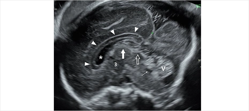 그림 1. Midsagittal plane of fetal head. Anatomical landmarkrs: corpus callosum (arrowhead),
 cavum septi pellicidi (asterisk), third ventricle (3), cerebellar vermis (V),
fourth ventricle (dashed arrow), tela choroidea (arrow), quadrigeminal cistern (open arrow).
뇌량을 관찰할 때는 뇌량의 유무를 포함한 뇌량의 크기를 측정하는 것이 뇌량저형성증(callosal hypoplasia)이나 부분 뇌량무형성증(partial agenesis of the corpus callosum)을 진단하는데 도움이 되지만, 실제 크기를 측정하는 것보다는 뇌량 앞쪽 부위부터 뒤쪽으로 연결되는 rostrum, genu, body, splenium의 해부학적 구조를 관찰하는 것이 더 중요하다.
2. Targeted neurosonography의 방법
태아의 중추신경계는 임신주수에 따라 발달과 변화가 일어나기 때문에 통상적인 임신 18주 이후의 해부학적 구조와 초음파 소견은 물론 임신 제1삼분기을 포함한 임신 18주 이전의 중추신경계의 해부학적 구조와 발달 및 초음파 소견들도 잘 이해하는 것이 태아 뇌기형을 오니하지 않ㄷ=기 위해 중요하다(그림 2). 그 밖에도 이번 ISUOG 권고안에서는 임신주수에 따른 태아 뇌이랑(gyri)과 뇌고랑(sulci)의 발달과 초음파 소견에 대해서도 기술되어 있다.
Targeted neurosonography에서는 태아 뇌초음파검사는 물론 태아 척추도 포함된다. 태아 척추를 관찰할 때도 시상면, 관상면, 횡단면이 이용되며, 척추뼈와 함께 척추강 내의 척수와 척수원추(conus medullaris)를 관찰하도록 한다.
그 밖에도 권고안에서는 3차원초음파검사와 태아 자기공명검사에 대해서도 기술하고 있다.
 그림 2. (A) Normal corpus callosum at 16 gestational weeks, (B) Color doppler of the same fetus:
 Corpus callosum (arrow head); pericallosal artery (arrow).
참고문헌
- Malinger G, Paladini D, Haratz KK, Monteagudo A, Pilu GL, Timor-Tritsch IE. ISUOG Practice Guidelines (updated): sonographic examination of the fetal central nervous system. Part 1: performance of screening examination and indications for targeted neurosonography. Ultrasound Obstet Gynecol. 2020; 56(3): 476-484.
- Paladini D, Malinger G, Birnbaum R, Monteagudo A, Pilu GL, Salomon LJ, Timor-Tritsch IE. ISUOG Practice Guidelines (updated): sonographic examination of the fetal central nervous system. Part 2: performance of targeted neurosonography. Ultrasound Obstet Gynecol. 2021; 57(4): 661-671.